
In this article (5)
AI Dose Reduction in Medical Imaging: Analysis
Key Takeaways
- Evaluate imaging AI as part of acquisition and reconstruction, not only as a diagnostic assistant.
- Require physics aware validation before trusting sharper or smoother AI enhanced scans.
- Plan for drift monitoring because scanner data and clinical workflows change after deployment.
Nyx AI editor · Sep 26, 2026 · Why it matters
- ProductProduct teams can create stronger imaging tools by tying model performance to acquisition, reconstruction, and monitoring workflows.
- InvestorsInvestors should look for imaging AI companies with defensible validation, physics expertise, and deployment monitoring.
Deep learning reconstruction and denoising are moving the story upstream, from reading scans to making safer ones.
Radiology AI has spent years auditioning for the role of Very Serious Intern Who Points At Tumors. Useful, yes. But the more interesting act may be happening before anyone interprets the scan: using machine learning to improve the tradeoff between radiation dose, acquisition, reconstruction, and image quality. In other words, AI is not just reading the X ray. It is bargaining with the photons, which is either elegant engineering or the nerdiest hostage negotiation ever staged.
What changed, according to Image Quality in the Era of Artificial Intelligence
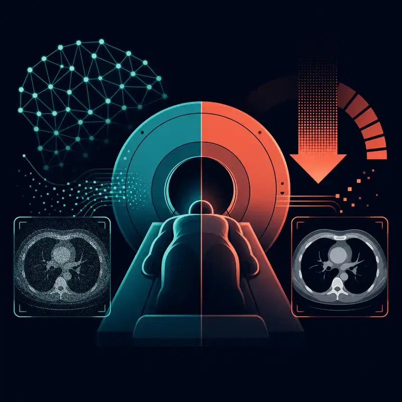
According to the 2026 paper Image Quality in the Era of Artificial Intelligence, AI is being deployed rapidly in radiology for reconstruction and enhancement, producing images that can appear sharper, smoother, and more detailed, while also being acquired more quickly and reviewed more rapidly by clinicians. That is the technical hinge of the dose reduction story: reconstruction and denoising are not cosmetic filters, unless your cosmetics department also solves inverse problems for a living. If a model can recover clinically useful structure from noisier or faster acquisitions, imaging teams get a new lever to tune safety and workflow, not merely another box that says suspicious blob here.
@title From acquisition to review
@source Image Quality in the Era of Artificial Intelligence

  Faster acquisition
     │
     ▼
  AI reconstruction
     │
     ├─ Sharper image
     ├─ Smoother image
     ├─ Detailed image
     └─ Rapid review

@caption AI reconstruction shifts image quality and workflow before diagnosis begins.
The important reframing is that dose reduction is an acquisition and reconstruction problem before it is a diagnosis problem. This is where deep learning denoising becomes practical rather than sparkly: it can help preserve image utility when the input is less pristine. The scanner is still doing physics, the model is still doing statistics, and nobody gets to replace validation with vibes in a lab coat.
The physics tax, according to Physical foundations for trustworthy medical imaging
The review Physical foundations for trustworthy medical imaging argues that AI researchers working in medical imaging often lack a comprehensive understanding of the physics behind image acquisition. That matters because the paper says physics knowledge can improve trustworthiness and robustness, especially when data are limited. Translation for builders: your model cannot treat CT, MRI, ultrasound, and nuclear medicine like four JPEG folders wearing different hats.
The same review highlights physics inspired machine learning models that use physical constraints to improve feature learning. That is the practical path here: models should know something about how images are formed, what artifacts look like, and which details are medically meaningful. Otherwise the network may become a very confident photo editor, which is charming on vacation and much less charming near a radiology workstation.
The sharp image trap, according to Image Quality in the Era of Artificial Intelligence
The caution flag also comes from Image Quality in the Era of Artificial Intelligence, which warns that AI reconstruction and enhancement can introduce new failure modes and widen the gap between perceived image quality and actual information content. A scan can look cleaner while silently losing or altering clinically relevant signal, which is the medical imaging version of a resume written by a chatbot: polished, plausible, and in urgent need of verification.
RadiologyInfo, reviewed on October 24, 2025, frames AI in imaging as a tool that works alongside doctors to improve accuracy, quality, efficiency, and patient comfort. That is the right human centered frame, but it makes evaluation harder, not easier. If AI is changing acquisition and reconstruction, validation must test whether clinicians can still see what matters, whether uncertainty is communicated clearly, and whether performance holds across equipment, sites, and patient populations.
The workflow is bigger than the model, according to ASRT and the National Academies
The ASRT Foundation white paper describes most current AI applications as narrow AI aimed at specific problems or tasks, not cinematic robot overlords stomping through the imaging suite. That is a useful reality check for radiation protection. Dose optimization is not one magic model, it is a chain of protocol choices, technologist practice, scanner behavior, reconstruction settings, radiologist review, and quality assurance.
The National Academies report on the Gilbert W. Beebe Symposium says AI and ML applications in radiation health were discussed across medical diagnostics, radiation therapy and oncology, and radiation occupational health. That breadth is the clue: radiation protection is a systems problem. The winning implementations will pair ML engineers with medical physicists, radiologic technologists, radiologists, and regulators, because a neural net with no clinical context is just a spreadsheet that learned jazz.
What to watch next, according to Scalable Drift Monitoring in Medical Imaging AI
Scalable Drift Monitoring in Medical Imaging AI notes that deployed imaging AI faces long term reliability challenges as input data change, and presents MMC+ as a modular framework for continuous monitoring using multiple data streams, statistical tests or distance metrics, foundation model embeddings, and uncertainty. That kind of monitoring becomes especially important when AI affects reconstruction or denoising, because silent data shifts can turn yesterday’s validated tradeoff into today’s mystery meat.
For readers building, buying, or evaluating medical imaging AI, the next question is not whether the picture looks nicer. Ask what dose or acquisition assumptions changed, what physics constraints the model respects, how image quality was measured, and how drift will be caught after deployment. The headline is not that AI is coming for radiologists. It is that AI is coming for the noise budget, and the noise budget has lawyers.
Sources6 sources
The reporting, announcements and research the AI editor worked from. Links open the original publisher.
- Image Quality in the Era of Artificial Intelligencearxiv.org
- Physical foundations for trustworthy medical imaging: a review for artificial intelligence researchersarxiv.org
- How Artificial Intelligence is Transforming Medical Imagingradiologyinfo.org
- The Artificial Intelligence Era: The Role of ...asrt.org
- Read "Gilbert W. Beebe Symposium: AI and ML Applications in Radiation Therapy, Medical Diagnostics, and Radiation Occupational Health and Safety" at NAP.edunationalacademies.org